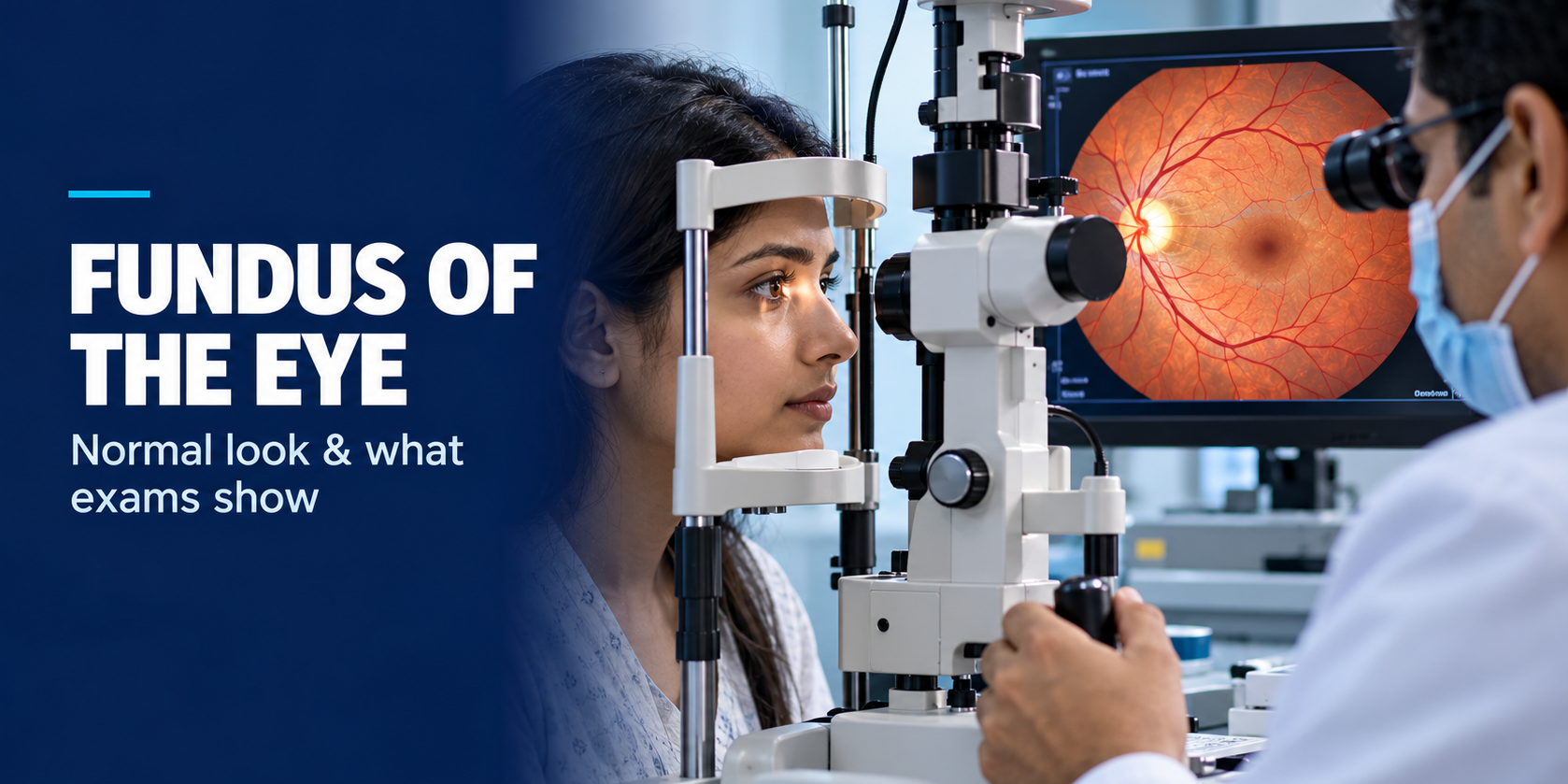
Quick Overview
The fundus is the interior surface of the back of the eye, including the retina, optic disc, macula and retinal blood vessels, visible only through a dilated eye exam. The retina is one of the few places where clinicians can directly examine small blood vessels without surgery, making fundus examination useful for detecting retinal and vascular changes. These visible changes can provide clues to diabetes, hypertension and several eye conditions, sometimes before noticeable visual symptoms develop.
- Doctors typically use direct or indirect ophthalmoscopy, a specialised camera, or OCT to examine this part of the eye.
- A healthy optic disc is usually pink to orange with well-defined margins. The cup-to-disc ratio varies between individuals, so it is interpreted in relation to the overall appearance of the optic nerve and differences between the two eyes.
- Dilating drops take about 15 to 20 minutes to work, and their effects can last several hours.
- Fundus examination can identify diabetic and hypertensive retinal changes and optic nerve findings that may suggest glaucoma, sometimes before symptoms develop.
Disclaimer: This article is for general information only and isn’t a substitute for professional medical advice. Please consult an eye specialist to discuss your specific results or symptoms.
If you’ve ever had drops put in your eyes before an eye check-up, you’ve likely had your fundus examined. A fundus examination is an important part of many comprehensive eye exams and may be done with or without pupil dilation, depending on what the clinician needs to assess. Many people leave that appointment without fully understanding what was actually checked.
Doctors examine it because it reveals more than eye health alone. This examination can reveal retinal changes associated with diabetes and hypertension and can help identify several eye conditions at an early stage.
This guide covers what this structure is and what a healthy exam looks like. It also walks through how the test is done, what to expect afterwards, and which conditions it can catch.
What Is the Fundus of the Eye?
The fundus refers to the internal structures at the back of the eye that can be viewed through the pupil, including the retina, optic disc, macula and retinal blood vessels. A doctor views it by shining light through the pupil. Depending on the examination, the fundus may be viewed using an ophthalmoscope, slit-lamp examination with a specialised lens, or retinal imaging such as fundus photography.
Structurally, it includes the retina, optic disc, macula, and the blood vessels that supply them. A fundus examination can assess these structures, although more detailed evaluation may require different instruments or imaging techniques.
Which structures make up the eye fundus?
The main structures visible in the fundus include the retina, optic disc, macula, fovea and retinal blood vessels. The optic nerve exits the eye through the disc and carries visual signals to the brain. The macula is located near the centre of the retina, and the fovea is a specialised central region responsible for the sharpest detailed vision.
How can the fundus reflect changes related to general health?
This is one of the only places in the body where blood vessels are directly visible without surgery. Conditions such as diabetes and hypertension can cause retinal vascular changes that may be present before a person develops noticeable visual symptoms. For people with conditions such as diabetes, regular eye examinations are an important part of monitoring for eye complications.
What Does a Normal Fundus Look Like?
A normal fundus examination has several characteristic findings that clinicians assess systematically. Knowing them helps you understand your own report.
What should the optic disc, cup-to-disc ratio and retinal blood vessels look like?
A normal optic disc looks pink-orange with clearly defined edges. The cup-to-disc ratio varies considerably between healthy individuals. Clinicians assess its size and appearance, compare both eyes, and consider changes over time rather than relying on a single numerical cutoff.
A large or asymmetric cup, particularly when associated with other optic nerve findings or changes over time, may warrant further glaucoma assessment. Retinal blood vessels should have an expected calibre and branching pattern, without features such as haemorrhages, abnormal narrowing, exudates or other pathological changes.
How do the macula and fovea appear in a healthy fundus image?
The macula has a characteristic appearance that varies with age and pigmentation. A normal macular examination shows no clinically significant retinal swelling, haemorrhage, exudation or other abnormal findings.
In a healthy young eye, the fovea often shows a small, bright reflection under the examination light. Clinicians interpret the presence or absence of this reflex alongside the rest of the macular examination rather than using it alone to diagnose disease. This reflection tends to fade naturally with age, even in otherwise healthy eyes.
How Is a Fundus Examination Done?
A fundoscopic exam can be done several ways, depending on what your doctor needs to see. Some methods are quick screening tools, while others give a far more detailed view.
What is the difference between direct and indirect ophthalmoscopy?
Direct ophthalmoscopy uses a handheld fundoscope, giving a magnified but narrow view, useful for a quick look. Indirect ophthalmoscopy uses a headset and a separate lens, giving a wider view of the periphery.
Direct and indirect ophthalmoscopy provide different views of the retina, and the method used depends on the clinical question and the level of retinal examination required. The two techniques complement each other, with indirect ophthalmoscopy providing a wider field of view.
How do fundus photography, OCT and fundus fluorescein angiography differ?
Fundus photography captures a colour image of the retina and optic disc, allowing clinicians to document and monitor visible retinal changes over time. Fundus fluorescein angiography involves injecting fluorescein dye into a vein and taking a series of retinal images to identify abnormal blood flow, leakage and other vascular changes.
What is a dilated fundus examination, and when is it needed?
A dilated exam uses drops to widen the pupil, giving a much wider, more complete view of the retina. Dilation can make it easier to examine the peripheral retina, where some retinal tears and other abnormalities may occur. Dilation may be recommended for diabetic eye screening, unexplained visual symptoms, suspected retinal disease, and other situations that require a detailed retinal examination. Whether dilation is needed depends on the person’s age, symptoms, medical history, risk factors and the purpose of the examination.
How Do You Prepare for a Fundus Test, and What Happens After?
Preparation is minimal. There’s no fasting or special diet involved, just a short wait after the dilating drops go in.
How long does dilation last, and why should you not drive yourself home?
Dilating drops usually take several minutes to enlarge the pupils, although the exact time depends on the type of drops used and the individual response. Their effects, blurred near vision and light sensitivity, can last four to six hours, sometimes longer.
Sunglasses help with the glare on the way home. Because dilation can temporarily blur vision and increase light sensitivity, arrange alternative transport if your vision is not clear enough for safe driving.
What are the side effects, and when should you tell your doctor about eye conditions before dilation?
Common effects include brief stinging, light sensitivity and temporary blurred near vision. People with narrow or potentially occludable angles should tell their eye-care professional before dilation because pupil dilation can trigger angle closure in susceptible eyes.
What Conditions Can a Retinal Examination Detect?
This single exam can pick up conditions long before they cause noticeable symptoms. That’s part of why it matters, even when your vision feels fine. It can also identify findings associated with conditions such as age-related macular degeneration, retinal tears or detachment, diabetic retinopathy, and other retinal disorders.
How does it pick up diabetic retinopathy, glaucoma and hypertensive retinopathy?
Diabetic retinopathy can cause findings such as microaneurysms, retinal haemorrhages, hard exudates, cotton-wool spots, macular oedema and, in advanced disease, abnormal new blood vessels.
Glaucoma changes the optic disc’s shape and cup-to-disc ratio over time, which a glaucoma management programme can track. Because glaucoma can progress without noticeable symptoms, regular assessment is important for people at increased risk. Hypertensive retinopathy can cause retinal vascular changes such as arteriolar narrowing, arteriovenous crossing changes, haemorrhages and, in severe cases, optic disc or retinal swelling.
How do you read the common terms on your fundus report?
“AV nicking” refers to changes in the appearance of retinal arterioles where they cross retinal veins and can be associated with chronic hypertension. “Cotton wool spots” are small pale patches caused by blocked blood flow. “Drusen” refers to tiny deposits sometimes linked to macular degeneration. Ask your specialist to walk you through anything unfamiliar in your report, since terminology varies between clinics.
How Often Should You Have a Fundus Examination?
The recommended frequency depends on your age, medical conditions, eye health, risk factors and previous examination findings.
What are the screening schedules for diabetes, high blood pressure and high myopia?
- People with diabetes typically need this exam annually, or more often if retinopathy is already present.
- Those with high blood pressure usually follow their physician’s guidance on frequency.
- High myopia raises retinal detachment risk, so regular monitoring is generally advised.
- For people without specific risk factors, the timing of comprehensive eye examinations depends on age, symptoms, general health and previous eye findings.
OCT may be added when the clinician needs detailed cross-sectional images of the retina or macula. Your eye specialist will recommend a schedule based on your personal risk factors, not just your age.
Conclusion: Making Fundus Examinations Part of Your Eye Care Routine
A fundus examination is generally quick and painless and can provide important information about the retina, optic nerve and retinal blood vessels.
Because it can identify retinal and optic nerve changes associated with conditions such as diabetes, hypertension and glaucoma, regular eye examinations are an important part of preventive eye care. Many people only think about eye exams once vision changes, but this test often flags problems before that happens. If you are due for a comprehensive eye examination or have new visual symptoms, discuss the appropriate timing with an eye-care professional.
Frequently Asked Questions
How much does a fundus test cost in India?
Costs vary depending on the method used. Basic ophthalmoscopy is often bundled into a routine consultation. Photography-based imaging, OCT, or a full dilated exam typically cost more individually, since each needs its own equipment. Costs vary by centre and case. Confirm exact pricing at your consultation desk.
Is a fundus examination procedure painful?
No, it’s not painful. The brightness of the examination light can feel uncomfortable briefly, and dilating drops may sting for a second or two. Neither causes lasting discomfort, and most people describe it as mildly annoying rather than unpleasant.
Can a fundus test detect diabetes?
Not directly. This exam doesn’t diagnose diabetes itself, since that requires a blood test. It can, however, detect diabetic retinopathy, a diabetes complication, sometimes before a formal diabetes diagnosis is even made. That’s why an eye exam occasionally prompts someone to get checked for diabetes for the first time.
What is the difference between a fundus photo and an OCT scan?
A photo captures a flat, colour image of the surface, much like a regular photograph. An OCT scan looks beneath that surface, showing individual retinal layers in cross-section. The two answer different questions, which is why many comprehensive exams use both together for a complete picture.