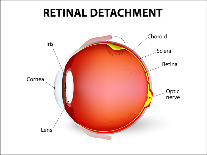
Retinal detachment describes an emergency condition. It is a condition wherein a thin layer of tissue (the retina) at the back of the eye pulls away from its usual position. It divides the retinal cells from the blood vessel layer that gives oxygen and nourishment. The more extended retinal detachment goes untouched, the higher your chances of permanent vision loss in the affected eye. Therefore, to avoid this situation, it is best to consult your doctor if there’s a need to go for Retinal detachment surgery.
Symptoms of Retinal Detachment
Retinal detachment itself is painless. But warning signs almost always appear before it occurs or has advanced, such as:
- The sudden appearance of many floaters. The specks that seem to drift through your field of vision
- Flashes of light in one or both eyes (photopsia)
- Blurred vision
- Gradually reduced side (peripheral) vision
- A curtain-like shadow over your visual field
Risk factors
The following factors multiply your risk of retinal detachment:
- Ageing — retinal detachment is more prevalent in people over age 50
- Previous retinal detachment in one eye
- Family history of retinal detachment
- Extreme nearsightedness (myopia)
- Previous eye surgery, such as cataract removal
- Past severe eye injury
- Early on eye disease or other disorders including retinoschisis, uveitis or thinning of the peripheral retina (lattice degeneration).
Diagnosis
Your doctor may exercise the following tests, instruments and procedures to identify retinal detachment:
Retinal examination: The doctor may use a tool that features bright light and special lenses. It can help examine the back of your eye, including the retina. This type of device provides a highly detailed view of your whole eye. Thus, it allows the doctor to see any retinal tears, holes or detachments.
Ultrasound imaging: Your doctor may utilise this test if bleeding has occurred in the eye, which makes it hard to see your retina.
Your doctor will examine both eyes even if you have symptoms in just one. If a tear is not diagnosed in this visit, your doctor may ask you to visit again within a few weeks. Only to make sure that your eye has not developed a delayed tear. It is still owing to the same vitreous separation. Also, if you undergo new symptoms, it’s essential to consult your doctor right away.
Treatment – Retinal detachment surgery
Surgery is most often used to restore a retinal tear, hole or detachment. For this, a variety of techniques are available. Ask your ophthalmologist regarding the risks and benefits of your treatment options. Above all, you can decide together on what procedure(s) is best for you.
Retinal tears
When a retinal tear or hole hasn’t yet advanced to detachment, your eye surgeon may advise one of the following procedures. It will check retinal detachment and also protects vision.
Laser surgery (photocoagulation) –Through the pupil, the surgeon directs a laser ray into the eye. As a result, the laser makes burns around the retinal tear, creating scarring that typically “welds” the retina to underlying tissue.
Freezing (cryopexy) – After administering a local anaesthetic to sedate your eye, the surgeon applies a freezing probe right over the tear to the eye’s outer surface. The freezing induces a scar that helps protect the retina to the eyewall.
Both of these procedures are on an outpatient basis. After the process, you’ll likely be advised to evade activities that might jar the eyes — such as running — for a week or two.
Coping and support
Retinal detachment may lead you to vision loss. Depending on the amount of vision loss, your lifestyle might vary accordingly.
As you learn to live with low vision, you may find the following ideas handy:
Get glasses –Optimise the vision you have with glasses. Use them as they are specifically tailored for your eyes. Also, ask for safety lenses to guard your better-seeing eye.
Brighten your home –Have a suitable light in your home for reading and other purposes.
Make your home safer –Remove throw rugs. Put coloured tape on the edges of steps. Consider setting up motion-activated lights.
Seek others help –Inform friends and family members of your vision issues.It will surely bring you some advice.
Get help from technology –Digital talking books and computer screen readers can assist with your reading.
Talk to others with impaired vision –Take help of online networks, support groups and resources for people with impaired vision.
When to see a doctor?
Seek direct medical attention if you are going through the signs or symptoms of retinal detachment. It is a medical emergency condition in which you can lose your vision for life.